L'esperienza pluridecennale in diagnostica ecografica incontra l'innovazione

Decades of experience in ultrasound diagnostics meets innovation

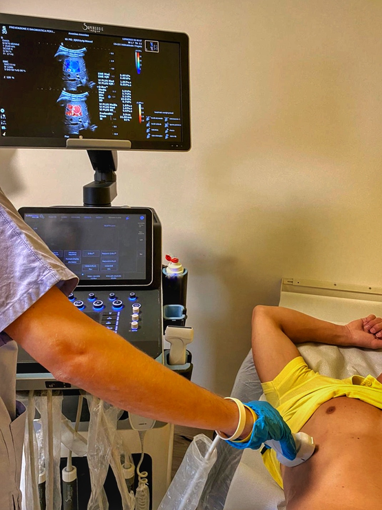
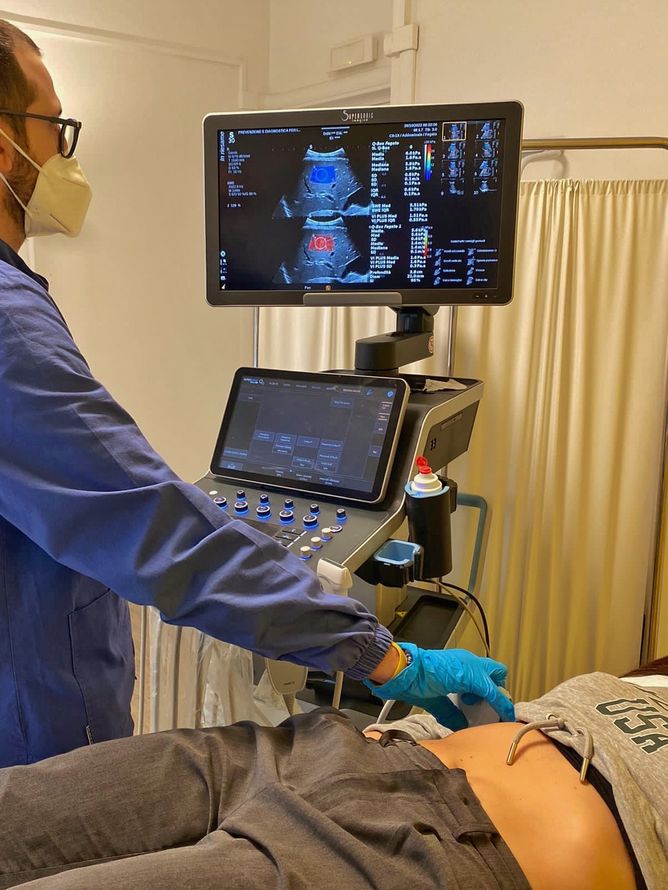
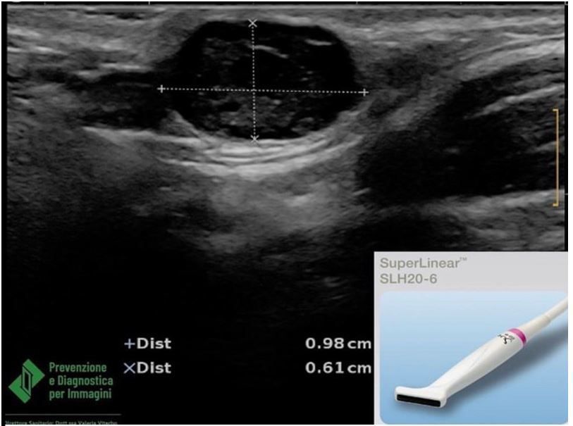
Prevenzione e Diagnostica per Immagini uses state-of-the-art equipment to perform breast, abdominal, thyroid, dermatological, musculoskeletal and soft tissue ultrasound scans. 


State-of-the-art equipment is used at the centre which also allows for the multi-parametric assessment of the liver with the Aixplorer® Mach system and the hepatic Shear Wave and mammary and thyroid nodules.

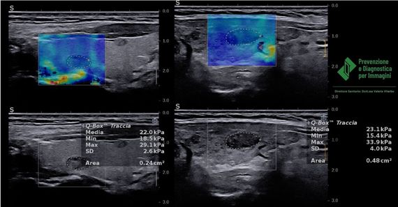
Shear Wave and Fibroscan


In patients with hepatopathy, Shear Wave Elastosonography makes it possible to assess the elasticity of the liver by calculating its 'stiffness', expressed in kiloPascals (kPa), to define the degree of liver fibrosis and correlate it to a numerical value that can be checked periodically, usually every year, to follow the evolution of the disease.


Recent scientific evidence shows that ShearWaveTM Elastography (SWE) compared to Fibroscan, which is its precursor, allows more accurate differentiation of the early stages of liver fibrosis and is therefore more effective than Fibroscan. Book your examination now with very short waiting times.

  • Ultrasound

    Slide title

    Write your caption here
    Button
  • Ultrasound investigation

    Slide title

    Write your caption here
    Button

Evaluation of hepatic steatosis


In the case of fatty liver, it is important to assess the degree of hepatic steatosis in order to carry out a precise and non-invasive staging of the disease and to be able to have a safe assessment of the progress of the disease at subsequent check-ups.


Indeed, we know that advanced steatosis is at the origin of more serious liver diseases such as fibrosis and cirrhosis.

With previous ultrasound scanners, the evaluation of  fatty liver was completely entrusted to a visual assessment performed by the sonographer and therefore an adequate quantification of steatosis was not possible.


Today, thanks to the Aixplorer® system, in use at our centre, it is possible to quantify the fat in the liver with precise numerical values that can be compared over time, such as the intrahepatic sound speed, attenuation coefficient and hepatic-renal index (B-Mode Ratio).

Quantifying the hardness of a suspected thyroid nodule: more information for the endocrinologist


Elastosonography makes it possible to obtain information on the hardness of a thyroid nodule.

The principle of this technique lies in the fact that the compression with the ultrasound probe of the tissue to be examined produces a distortion, less for hard tissue and more for soft tissue. This hardness can be detected by an ultrasound scanner using dedicated software by quantifying the 𝗦𝘁𝗶𝗳𝗳𝗻𝗲𝘀𝘀 values, expressed in kiloPascals (kPa).


Malignant thyroid nodules usually present greater hardness compared to normal tissue and benign nodules.

For these reasons, elastosonography is one of the best non-invasive tools to estimate the risk of malignancy of the nodular lesion and thus reduce the use of thyroid needle aspiration.


This method can also be used for those nodules that, subjected to needle aspiration, have an uncertain cytological diagnosis. In our centre, thanks to the Aixplorer® system, in addition to assessing the morphological,

ecostructural and vascularisation characteristics of thyroid nodules, as in a standard Echo-Colour-Doppler examination, we also perform the elastosonographic study of suspicious nodular lesions (Shear Wave).

  • Ultrasound

    Slide title

    Write your caption here
    Button

Quantificare la durezza del nodulo tiroideo sospetto: un'informazione in più per l'endocrinologo


𝙇’𝙚𝙡𝙖𝙨𝙩𝙤𝙨𝙤𝙣𝙤𝙜𝙧𝙖𝙛𝙞𝙖 consente di ottenere informazioni sulla durezza di un nodulo tiroideo.

Il principio di tale tecnica risiede nel fatto che la compressione con la sonda ecografica del tessuto da esaminare, produce una distorsione, minore per i tessuti duri e maggiore per i tessuti soffici; questa durezza può essere rilevata dall’ecografo tramite un software dedicato quantificando i valori di “𝗦𝘁𝗶𝗳𝗳𝗻𝗲𝘀𝘀”, espressi in kiloPascal (kPa).

I noduli tiroidei maligni presentano, di solito, una 𝗱𝘂𝗿𝗲𝘇𝘇𝗮 𝗺𝗮𝗴𝗴𝗶𝗼𝗿𝗲 rispetto al tessuto normale e ai noduli benigni.

Per questi motivi 𝗹’𝗲𝗹𝗮𝘀𝘁𝗼𝘀𝗼𝗻𝗼𝗴𝗿𝗮𝗳𝗶𝗮 è uno dei migliori strumenti, non invasivi, per stimare il rischio di malignità della lesione nodulare e pertanto ridurre il ricorso agli agoaspirati tiroidei. Questa metodica può essere utilizzata anche per quei noduli che, sottoposti ad ago aspirato, hanno una diagnosi citologica incerta. Nel nostro centro, grazie al sistema Aixplorer®, oltre a valutare le caratteristiche morfologiche, ecostrutturali e di vascolarizzazione dei noduli tirodei, come accade in un esame Eco-Color-Doppler standard, effettuiamo lo studio elastosonografico delle lesioni nodulari sospette (la Shear Wave).

  • Ecografia

    Slide title

    Write your caption here
    Button
  • Titolo diapositiva

    Scrivi qui la tua didascalia
    Pulsante

Dermatological ultrasound


To assess skin and subcutaneous pathologies, it is necessary to use high-resolution and high-frequency ultrasound probes. In dermatological ultrasound, probes with a frequency of 13 to 50 Mhz (MegaHertz) are used. The higher the frequency, the lower the ultrasound penetration into the skin tissue and the better the evaluation of superficial tissue. Therefore, only with dedicated probes is it possible to distinguish in ultrasound the three layers that make up the skin: epidermis, dermis and underlying tissue. At our clinic, we perform dermatological ultrasound with a dedicated high-frequency probe (6 to 20 Mhz).

  • Titolo diapositiva

    Scrivi qui la tua didascalia
    Pulsante

Ecografia dermatologica


Per valutare le patologie cutanee e sottocutanee è necessario utilizzare sonde ecografiche ad alta risoluzione e ad alta frequenza. Nell'ecografia dermatologica, infatti, si utilizzano sonde con frequenza da 13 a 50 Mhz (MegaHertz). Tanto maggiore è la frequenza, tanto più è ridotta la penetrazione degli ultrasuoni nel tessuto cutaneo e migliore la valutazione dei tessuti superficiali. Pertanto solo con sonde dedicate è possibile distinguere in ecografia i tre strati che costituiscono la cute: epidermide, derma e tessuto sottostante. Nel nostro centro eseguiamo ecografia dermatologica con sonda dedicata ad alta frequenza (da 6 a 20 Mhz).

Call for more information

Call the clinic
Share by: